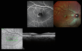
Spotlight Case: Early Diagnosis of MacTel Type 2 Using Multimodal Imaging with SPECTRALIS
A 28-year-old male patient presented with visual distortion in his right eye. Leveraging Heidelberg Engineering’s SPECTRALIS multimodal imaging—comprising OCT, Multicolor Imaging, and Fluorescein Angiography—clinicians successfully diagnosed Macular Telangiectasia Type 2 (MacTel Type 2) at an early stage. The case exemplifies how subtle pathological changes can be detected well before classic signs appear, ensuring timely and precise intervention.
Diagnosing MacTel Type 2 Using Heidelberg Multimodal Imaging
A Case Study Highlighting the Role of OCT, Multicolor Imaging, and FFA
Overview
Macular Telangiectasia Type 2 (MacTel Type 2) is a rare retinal disease that affects the parafoveal region, leading to progressive vision loss. Early diagnosis is critical, and Heidelberg Engineering’s advanced multimodal imaging plays a pivotal role in detecting subclinical features of this condition.
Patient Presentation
A 28-year-old male presented with visual distortion in his right eye for three months.
-
BCVA: 6/9 (Right), 6/6 (Left)
-
Fundus Exam: Loss of parafoveal retinal transparency
-
Anterior Segment: Normal
Multimodal Imaging Findings
OCT
-
Revealed hyperreflective lesions in the parafoveal area
-
Indicated early structural disruptions associated with MacTel Type 2
Multicolor Imaging (MC)
-
Detected focal zones of blood leakage
-
Identified vascular changes crossing the foveal avascular zone (FAZ)
-
Provided superior detail of subtle anomalies
Fluorescein Angiography (FFA)
-
Early phase: Unremarkable
-
Late phase: Capillary leakage visible, aligning with MC findings
Clinical Insights
Multimodal imaging enabled early, non-invasive detection of MacTel Type 2, even before clear signs were visible on traditional FFA. The combination of MC and OCT allowed for accurate diagnosis and provided a clear roadmap for monitoring progression.
Outcome & Follow-Up
-
Recommended regular imaging follow-ups
-
Lifestyle and dietary adjustments advised
-
Anti-VEGF therapy considered for future intervention if needed
Conclusion
This case underscores the transformative potential of Heidelberg’s multimodal imaging in diagnosing retinal diseases like MacTel Type 2. With its ability to reveal early vascular and structural changes, clinicians can intervene earlier and manage disease progression more effectively.